Cones
are sensitive to photopic higher levels of light,
especially hues, or colors. When entering a light
area from a dark one, we experience light adaption,
but cones responding to the higher light levels recover faster than
rods. The receptive photopigments in cones are called iodopsins.
Color
vision is most precise at the fovea, a small area
behind the iris and lens, which is what we use when we look directly
at an object in medium to high light levels. The fovea consists
entirely of cones, which makes it difficult to see an object in
low light settings by looking directly at it.
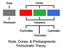 There
are believed to be three types of cones, each sensitive to a different
bandwidth of light and range of colors, and each containing a different
type of photopigment. Cones containing erythrolabe
are sensitive to long or red wavelengths, those containing chlorolabe
respond to medium or green wavelengths, and those containing cyanolabe
are affected by short or blue wavelengths. There
are believed to be three types of cones, each sensitive to a different
bandwidth of light and range of colors, and each containing a different
type of photopigment. Cones containing erythrolabe
are sensitive to long or red wavelengths, those containing chlorolabe
respond to medium or green wavelengths, and those containing cyanolabe
are affected by short or blue wavelengths.
This
theory of three types of cones is called trichromatic theory,
which proposes that each type of cone is genetically coded for receiving
and sending its specific color bandwidth to the brain.
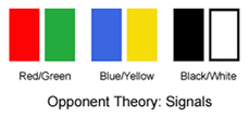 The
opponent theory proposes that the signals from
the retinal cones are further converted in the brain to sets of
opposing signals: red/green, blue/yellow, and black/white, which
are thought to genetically define the ways in which the human brain
processes color. Subjects in tests typically do not describe a color
as reddish green, or bluish yellow, or darkish light, indicating
that each of these combinations are processed as unique pairs of
opposites, and are not perceived in the brain to describe a single
color. The
opponent theory proposes that the signals from
the retinal cones are further converted in the brain to sets of
opposing signals: red/green, blue/yellow, and black/white, which
are thought to genetically define the ways in which the human brain
processes color. Subjects in tests typically do not describe a color
as reddish green, or bluish yellow, or darkish light, indicating
that each of these combinations are processed as unique pairs of
opposites, and are not perceived in the brain to describe a single
color.
|