| |
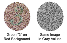 While
no two individuals can be expected to have exactly the same color
experience at any given time when viewing a specific scene, as many
as 10% of human males, and .05% of females, perceive colors much
differently than the average population. These differences are called
color vision deficiencies, and can range from a
slight shift from the norm in color perception to perceiving no
colors at all, or only perceiving gray values. Visual tests such
as the one depicted here are used to evaluate color deficiencies. While
no two individuals can be expected to have exactly the same color
experience at any given time when viewing a specific scene, as many
as 10% of human males, and .05% of females, perceive colors much
differently than the average population. These differences are called
color vision deficiencies, and can range from a
slight shift from the norm in color perception to perceiving no
colors at all, or only perceiving gray values. Visual tests such
as the one depicted here are used to evaluate color deficiencies.
Color
deficiencies in humans are relatively rare, and most affect males.
This is because inherited color perception is passed genetically
through the X chromosome, and normal females have two X chromosomes,
so the chances of both carrying a color deficiency is rare. Males,
however, have a greater chance of inheriting a color deficiency
through their single X chromosome, which is passed from the mother.
An
estimated 5% of males are anomalous trichromats,
those who can see all colors but have deficiencies in one or more
photopigments, causing color perception to shift away from the norm.
A dichromat
lacks one of the 3 cone photopigments, visually experiencing the
world without one of the red, green, or blue stimulations, or with
it markedly shifted from the average population. A dichromat lacking
erythrolabe, or the photopigment to see red, is
known as a protanope or an individual having protanopia.
One who lacks chlorolabe, the green photopigment,
is called a deuteranope and is affected by deuteranopia.
These red/green deficiencies are the most common dichromatic deficiencies,
and are usually inherited, affecting about 2% of males and .4% of
females.
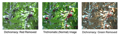
While
it's difficult to know how others with color deficiencies see the
world, the images below show the same scene (of red cherries in
green leaves) with red colors removed (left) and with green colors
removed (right).
The
rarest dichromat is the tritanope, who lacks cyanolabe,
or the photopigment to receive the color blue. This deficiency,
known as tritanopia, is more often acquired than
inherited, sometimes by disease or injury. Tritanopia affects less
than .1% of the population. A tritan shift can also be caused by
cataracts, or yellowing of the cornea caused by exposure to UV rays
or by aging.
The
most extreme example of color deficiency is the monochromat,
who sees the world with only one photopigment. An achromat,
also known as a rod monochromat, perceives differences
between light and dark gray values but does not see colors. A cone
monochromat sees with only one set of cones, perceiving
images only in red, green, or blue ranges. Monochromacy is rare,
affecting less than .01 % of the population.
We
all perceive color differently, but a small percentage of us perceive
it markedly differently than most. However, since we are all only
aware of own color perception, it's difficult to describe how one
person sees color versus another. And many people with color deficiencies
have leaned to identify colors using other cues, such as relative
lightness and darkness values, combined with shapes of items as
when comparing a banana and a tomato. The next modules in this program
can help in designing for everyone, in a way that doesn't include
identical values that are directly aligned with one another.
Click
Next to take the Biology of Color Quiz!
|
|